Updated Intramural Ectopic Ureter
Graphic courtesy Shalini Radhakrishnan/VIN
What is it?
An ectopic ureter is a birth defect in which one or both ureters in the kidneys connect to an abnormal location. This defect leads to the most common complaint: difficulty in house training and the pet leaking pee (urine). Ectopic ureters cause involuntary dribbling of pee, uncontrollable need to pee (urination), and urinary tract infections. Ongoing urinary tract infections can be fatal if they reach the kidneys, so it is important to diagnose and correct the ureter placement through surgery.
Normal Anatomy
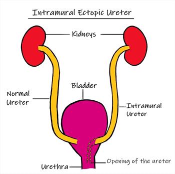
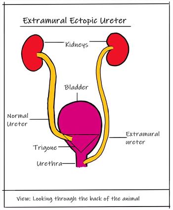
Each animal has two kidneys, which make urine. Each kidney has a collection tube, called the ureter, that connects to the bladder at a location named the trigone. Urine is emptied from the bladder through a single tube called the urethra. Therefore, urine moves from the kidneys, through the ureters, and into the urinary bladder, where the urine is stored until the pet purposely pees. When urination occurs, urine empties through the urethra outside the body.
Two Types
There are two types of ectopic ureters based on where these collection tubes connect inside the pet's body. Ectopic ureters do not connect and open at the bladder trigone, as they do in a normal animal.
Intramural ectopic ureter:
This type is the most common type in dogs. The collection tubes attach to the bladder at the trigone but then tunnel through the bladder wall, finally opening downstream from the bladder (i.e., the urethra).
Updated extramural ectopic ureter
Graphic courtesy Shalini Radhakrishnan/VIN
Extramural ectopic ureter:
This type is rare in dogs but more common in cats. This is when the ureters pass the bladder and connect and open downstream from the bladder. This connection can be further along the urinary tract or even to the reproductive tract (i.e., urethra or vagina).
Who Gets it?
An ectopic ureter is an abnormality that occurs while the fetus is developing. It is a rare abnormality in dogs, and even less common in cats. It is most commonly seen in young pets because that is the age when signs are first noticed.
Cats
- Sex: Both females and males equally
- Breeds: Himalayan, Persian, Maine Coon
- Age: Mostly young pets
Dogs
- Sex: Females nine times more likely than males
- Breeds: Labrador Retriever, Golden Retriever, Siberian Husky, West Highland White Terrier, Miniature Poodle, Newfoundland, and Soft Coated Wheaten Terriers
- Age: Mostly young pets
What are the signs?
- Urine leakage or dribbling (incontinence)
- Hard to house train; lots of accidents
- Urinary tract infection, more common in females
- Licking of genital area
- Rash on the genital area
- Urine staining and genital area is persistently wet
- Male dogs can have fewer signs and develop signs of urinary incontinence later in life.
- Not all cats and dogs will show signs.
How is it Diagnosed?
A complete physical exam and basic diagnostic tests, such as a complete blood count, blood chemistry, and urine analysis and culture will be done first.
Diagnosis requires imaging. The method chosen depends on availability, training of the person doing the imaging, and your animal’s specific case. There is no one imaging method that is the best, and multiple methods may be needed.
Radiographs and ultrasound are two imaging methods that show the kidney shape and size.
Contrast CT and cystoscopy are two methods that are gaining popularity as they give a better visual of the ureters. Cystoscopy is a procedure where a small camera is placed at the pet’s urethral opening and goes into the body looking for the ureter openings. It gives the veterinarian a great visual of where the abnormality is located.
The type of ectopic ureter, location, and size have no effect on the prognosis and outcome of treatment. However, imaging is necessary to determine which surgical procedure to perform and the surgical approach the veterinarian will use.
- Radiographs are usually readily available and cost-effective, but have limited ability to identify specifics.
- Ultrasounds are non-invasive, visualize much of the urinary tract, and are relatively readily available. Your veterinarian may be able to diagnose ectopic ureters with ultrasounds, but it may be difficult to visualize the lower urinary tract
- CT scans provide a good visual of the entire urinary tract but are expensive and not always available
- Cystoscopy provide a good visual of the entire lower urinary tract and sometimes a repair can be done right then. It is not always available.
What is the Surgery and Treatment?
The type of surgery used to correct the misplaced ureter depends on its location. Studies show that there is no significant increase in risk or complications between any of the surgeries. All the surgical options have similar long-term outcomes.
Ureteroneocystostomy
This procedure forms a new ureter and bladder opening. The surgery fixes the extramural type by creating a new bladder opening for the ureter in the correct spot. This surgery may cause swelling of the ureter during healing, but it will resolve in a few weeks.
Ureteronephrectomy
This procedure removes the ureter and kidney. The surgery is done on patients that also have a nonfunctional or chronically infected kidney attached to it. However, it can be difficult to tell if a questionable kidney is not functioning.
Laser Cystoscopy
This surgery is a new method specific to treating the intramural type. The laser cuts a new ureter opening into the bladder at the normal area. Surgery is less expensive because it can be done right after the initial cystoscopic diagnosis, while the patient is still under general anesthesia.
What is the Aftercare and Long-term Outcome?
Depending on the type of surgery, the aftercare and time of recovery may vary. The main post-surgical problems are continued uncontrolled peeing, leakage of pee into the body, swelling at the surgical site, and urinary tract infection. Studies show uncontrolled peeing resolves in cats that get a ureteronephrectomy. For dogs undergoing any of the surgeries for the birth defect, almost half will still have urinary leakage. If this occurs, additional medications will be needed for the rest of the pet’s life to help control urination.
It is not clear why some animals have continued urine leakage after surgery. Pets affected with ectopic ureters should not be bred.