Mass Aug 2017
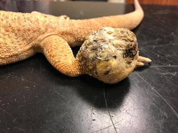
A mass on the leg of a bearded dragon that is very characteristic of an abscess. Photo by Dr. Jessica Peace.
Abscesses in reptiles are formed by accumulations of trapped white blood cells. This trapping is called encapsulation because the white blood cells are contained in a capsule of tissue. This encapsulation of the white blood cell material typically causes firm swellings or masses to form. In mammals — like you and me — there are enzymes that break down the white blood cells and turn them into a liquid, such as the pus in a pimple or ant bite. However, reptiles do not have these enzymes and so the abscesses are solid and contain a cheese-like pus and because of this and the presence of a fibrous capsule, the term fibrescesses is sometimes used.
Abscesses are commonly seen on the skin or often felt just under it but they can develop anywhere in or on the body. Usually an infectious agent causes the abscess to form. The most common causative organisms are Gram negative bacteria, so called because they do not stain with a common stain used to identify types of bacteria. There are many different chemicals that are used to help identify bacteria and other organisms; these are called stains. Different types of organisms will stain different colors, or some like gram-negative bacteria do not take up any color at all. However, other types of bacteria, such as anaerobic and acid-fast can also cause abscesses in reptiles. In addition, other organisms such as fungi and parasites (protozoa, nematodes, cestodes), foreign material such as plants, glass, fiberglass, plastic, and wood can also create abscesses. Recently, a new or novel Actinomyces species (a Gram-positive bacteria) has been shown to be the cause of cloacal and hemepene abscesses in breeding ball pythons and a fungus (Emydomyces testavorans) is an emerging cause of shell and internal abscesses in fresh water turtle species.
What may lead you to suspect your reptile may have an abscess? Abscesses can form almost anywhere on, or inside the body, so the signs you see depend on where it is and what organ it is affecting.
For abscesses on the skin or just under the skin, called cutaneous and subcutaneous respectively, you will often see some of the following signs:
- Lump(s), swellings, masses, and/or the two sides of the body having a different shape.
- Injury to the skin over the mass may or may not be evident.
- The reptile may be lame or unable to walk.
- Nonspecific signs, including depression, inactivity, and/or anorexia
- Bite wounds or trauma from a cagemate or other animal in the house such as a cat or dog.
- Common sites:
- Head (especially along upper or lower jaw bones and nostril area)
- Neck
- Legs, feet, toes
- Over the spine or the tail
For those abscesses that form inside the body:
- Nonspecific, including depression, inactivity, and/or anorexia (e.g., hepatic abscesses)
- Anorexia with abscesses located in the mouth, body cavity, or gastrointestinal tract
- Specific to internal organs involved, for example:
- Breathing problems with lung abscesses
- Diarrhea, darkened stools, blood in stools or constipation from gastrointestinal abscesses
- Loss of balance, seizures from abscesses in the brain
Abscesses in the mouth cavity:
- Loss of tissue around the oral cavity (especially toward the front of the head), exposure of oral mucus membranes and bone
- Abscessation along gum line may occur with periodontal disease in lizards with acrodont dentition
- Corner of the mouth:
- Asymmetry at corners of mouth, inability to close mouth
- Associated with gland in Old World chameleons (especially Jackson’s chameleons)
Sometimes the abscess will be located in a specific area, including:
- Aural (ear) abscesses; signs include:
- Anorexia (lack of appetite)
- Swelling on side of head
- Asymmetry to head (unilateral); one side is larger than the other
- Entire head enlarged (bilateral)
- Head tilt
- Common in box turtles and aquatic turtles
- Hemipenal abscesses/scent gland abscesses; signs include:
- Swelling associated with cloacal/vent area
- Fecal and urate accumulation around vent
- Blood from cloacal area seen
- Odor from around vent
- Anorexia
- Loss of breeding interest
- Scent gland abscesses (snakes):
- Swelling just cranial, adjacent, and caudal to vent
- Asymmetric when unilateral (may be bilateral)
- Associated with possible odor
- May be concurrent with hemipene involvement
- Subspectacular (under the spectacle of the eye) abscesses; signs include:
- Enlarged, swollen, nonsymmetrical (if unilateral)- one eye appears larger than the other
- The eye is no longer clear but instead cloudy, white, or yellow
- Owner suspects loss of vision.
- Change in eye(s)
- May be associated with an infection in mouth
- Anorexia
It is important for your veterinarian to determine the cause. In some cases, abscesses are associated with or caused by bacteria such as Salmonella or Mycobacteria that can cause disease in humans.
Affected Reptiles
Abscesses are common in all reptiles. There appears to be an increased incidence in juvenile reptiles due to:
- Demands of growth that require more exacting husbandry that is often lacking
- A developing immune system (immune suppression with inappropriate husbandry)
- Housed in pairs or groups, resulting in increased cagemate aggression/stress
- Male reptiles commonly develop hemipene or copulatory organ abscesses.
Active or highly stressed reptiles that do not acclimate to captive life are predisposed to abscesses. Examples include:
- Reticulated pythons, Asian water dragons and Basilisks (abscesses at front of head secondary to trauma from escape attempts)
- Old World chameleons (feet/toes, glands at the corners of the mouth)
Some common risk factors that may lead to abscesses include:
- Poor husbandry/stress (often resulting in immune suppression):
- Incorrect temperatures
- Lack of or too much moisture/humidity
- Incorrect caging (small size, lack of visual barriers/hide areas)
- Overcrowding
- Malnutrition (hypovitaminosis A)
- Trauma:
- Prey items (rodents, insects)
- Cagemate interactions (bites and scratches)
- Other house pets
- From contact with cage or cage furniture (fiberglass, sharp edges)
Diagnosis
To diagnose an abscess, your veterinarian will begin with a thorough husbandry history and physical examination. There are a wide variety of conditions that can mimic or are similar to the signs caused by an abscess that include:
- Fractures
- Metabolic bone disease (nutritional secondary hyperparathyroidism [NSHP])
- Neoplasia (cancer)
- Parasites (protozoa, cestodes, nematodes)
- Hematoma/pseudoaneurysm
- Mycobacteria
- Gout/pseudogout
- Sebaceous cysts
- Dermatophilosis (see Dermatophilosis [Rain Rot])
- Hypovitaminosis A results in squamous metaplasia involving secondary bacteria and abscesses
- For subspectacular abscesses:
- Pseudobuphthalmos (blocked nasolacrimal duct)
- Pseudobuphthalmos associated with flagellates (intestinal protozoa)
- Retained spectacle
While your veterinarian will often be able to diagnose an abscess just during a physical examination, some additional testing may be needed to obtain a diagnosis. This testing may be performed in the office, or samples may need to be sent out to a diagnostic laboratory.
Your veterinarian may obtain some material from a lump or bump using a syringe and needle; this is called a fine needle aspirate. An alternative is that a glass or plastic slide may be used to obtain some material for staining; this is called an impression smear. The material will be stained in the clinic and looked at under the microscope. The cells may show that there is an abscess. In addition, some of this material may be sent to a diagnostic laboratory for a culture and sensitivity to determine what type of bacteria is involved and to determine which antibiotic will be effective. A blood sample may be taken to see if the reptile has an infection and to determine if one of the organs is affected by an internal abscess.
A common diagnostic method that your veterinarian may suggest is a biopsy. This material is then sent out to a laboratory to look at the tissue and determine if there are any organisms or disease agents. This is called histopathological evolution, or histopath (some people call it a path report, even though that is not technically the correct name). Ultrasound and/or the use of an endoscope may be suggested by your veterinarian to help show the abscess and obtain the biopsy sample.
Radiographs (X-rays) can help show internal abscesses or bone destruction from the abscess. In some complicated cases, computed tomography (CT) or magnetic resonance imaging (MRI) may be suggested.
Treatment
In all cases, your veterinarian will review the husbandry history with you and make suggestions on improvements.
For the initial treatment, depending on what the physical examination reveals, your veterinarian will likely warm the patient to the upper end of the reptile’s preferred optimal temperature. For many commonly kept reptiles, this is between 80°F and 90°F (26°C and 32°C) and begin fluid therapy if needed. While reptiles sometimes get fluid therapy from an IV bag, more often it’s sub-cutaneous, in the body cavity, or in the bone (intraosseous).
In the majority of cases, your veterinarian will suggest that the abscess be surgically removed and then prescribe a course of appropriate antibiotics. If the abscess cannot be fully removed, aggressive surgical debridement (cleaning out the abscess) is needed. Both of these procedures will require anesthesia and pain medication. Flushing with a variety of solutions for one or more weeks after surgery is important. Your veterinarian will discuss that with you.
Most abscesses require systemic antibiotic therapy. Once the reptile is rehydrated and has an appropriate core body temperature, antibiotic therapy can be started. It is critical that you give the antibiotics exactly how your veterinarian describes and for the complete number of days; you should never stop the antibiotics without first talking with your veterinarian.
In all cases, a follow-up visit is recommended once the medication course is near completion to decide whether longer treatment may be necessary. In cases that require flushing of the open abscess area, healing should be rechecked before complete closure of the surgery site to make sure that the infection has cleared. Cases with bone involvement should be rechecked frequently during treatment with follow-up x-rays to assess progress with any bone change.
Prognosis and Prevention
Isolated abscesses that do not involve bone and that get early, aggressive treatment usually have a favorable prognosis. Cases that have a more guarded or a poor prognosis owing to possible sepsis (a body-wide condition resulting from bacteria in an infection) include those patients that have internal abscesses, debilitated patients with multiple abscesses, and patients that have bone involvement.
Recurrence may be seen in cases where the abscess is not able to be removed entirely. If the surgical incision closes prematurely, the abscess can re-form. This is especially true with deeper abscesses or those with concurrent bone involvement; internal abscesses or abscesses involving active glandular tissue such as scent gland abscesses; or abscesses involving the glands at the corner of the mouth in chameleons.
Prevention always involves correcting any underlying predisposing conditions and risk factors such as inappropriate husbandry and social stress, cage mate aggression and/or overcrowding that may lead to immune suppression. With hemipene abscesses and scent gland abscesses in breeding snakes, avoid trying to breed one male with too many females and keep the substrate clean during active breeding.
References for the two new emerging causes of abscesses
Oral, Cloacal, and Hemipenal Actinomycosis in Captive Ball Pythons (Python regius).
Language: English
Front Vet Sci. January 2020;7(0):594600.
Steven B Tillis 1, Marley E Iredale 1, April L Childress 1, Erin A Graham 1, James F. X. Wellehan 1, Ramiro Isaza 2, Robert J Ossiboff 1
Shell Lesions Associated With Emydomyces testavorans Infection in Freshwater Aquatic Turtles.
Language: English
Vet Pathol. May 2021;58(3):578-586.
Daniel B Woodburn 1, Michael J Kinsel 1, Caryn P. Poll 2, Jennifer N. Langan 3, Katherine Haman 4, Kathryn C Gamble 5, Carol Maddox 3, Albert B Jeon 6, James F. X. Wellehan 6, Robert J Ossiboff 6, Matthew C Allender 3, Karen A Terio 1