Stephanie Cruz-Rincon, DVM; Kathleen Freeman, DVM, PhD, DipECVCP, FRCPath, MRCVS
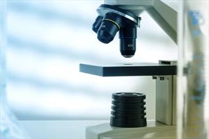
Microscope
What Is Histopathology?
Histopathology is the microscopic examination of stained tissues to look for a potential cause or the size of a diseased area. Staining is the process that applies dyes to the slide to highlight cells and make them easier to see.
What Is a Biopsy?
A biopsy is the removal and/or examination of cells or tissue from the body to figure out a disease's presence, cause, or extent.
Benefits of Histopathology
Histopathology can be a powerful diagnostic tool. There are many diseases that cause vague and nonspecific clinical signs. The results of histopathology can help your veterinarian know the specific disease your pet is suffering from and the required treatment.
In some cases, histopathology is the only way to find out for sure what condition your pet may have. Many diseases require histopathology to be confirmed. Some of these are various inflammatory, congenital (a condition existing from birth), and cancerous diseases.
Examples of inflammatory diseases that require histopathology to be diagnosed include inflammatory bowel disease, glomerulonephritis (inflammation of the glomeruli, which are part of the kidneys), and hepatitis (inflammation of the liver).
Unusual or severe skin abnormalities are instances when your veterinarian may initially recommend histopathology when making a diagnosis. Histopathology can be useful in a pet who isn’t displaying obvious signs of sickness. Some veterinarians may recommend taking biopsies of masses based on their size, location, duration, appearance, or if seemingly painful.
Cancer is a type of disease that often requires histopathology for diagnosis. Histopathology is often needed to correctly identify the mass as cancerous or not. This identification is important for detecting your pet's cancer and deciding the best treatment plan.
Biopsy Procedure
A tissue biopsy is obtained by removing a small piece of tissue. How that tissue is taken depends on the type of tissue and where that tissue is located.
Some methods of obtaining a biopsy do not require invasive surgery. Your pet may only need local anesthesia (numbing the area) and sedation in these cases. For example some skin lesions are often investigated with a simple skin punch biopsy.
Other biopsy methods may require general anesthesia, where your pet is “put under” and unconscious for the procedure. This option is usually for larger superficial masses or internal biopsies, like those of the intestines, and may involve the use of an endoscope (a small camera on a long flexible wand to see within the body) or exploratory surgery, where a body cavity like the chest or abdomen is opened to look for masses or other abnormalities.
Your veterinarian will explain how they will get a biopsy sample and decide whether your pet is stable enough to undergo general anesthesia if needed. Depending on the procedure, your veterinarian may also refer you to a veterinary specialist to obtain the biopsy and to help recommend further treatment.
Once a biopsy is obtained, it is sent out to a pathologist. A veterinary pathologist will examine or “read,” the tissue sample and report their findings. In most cases, the biopsy sample will take some time to ship to a laboratory and to be evaluated. Your veterinarian will give you an estimate of when to expect results.
Recovery
Your pet will have a wound where the tissue was removed and may have stitches. Your veterinarian should provide you with post-surgical instructions and may provide pain relief medications as needed.
The biopsy site needs to be kept clean and dry; your pet should not be able to lick it or irritate the wound. Your veterinarian may recommend an Elizabethan collar (cone) to help prevent your pet from touching it. If you have questions at any time about how your pet is healing, always contact your veterinarian.
Limitations of Histopathology
While histopathology can provide valuable information, it has limitations too. It helps your vet to find the diagnosis most of the time. Errors can happen when tissue sampling is not good enough (e.g., the biopsy was not representative of the affected area) or if there is a lack of clinical history. Your veterinarian can prevent many of these errors by collecting and handling the tissue appropriately and working closely with the pathologist.
Your veterinarian may request a second opinion on the biopsy if the results come back inconclusive or are unexpected, and they may recommend repeating a biopsy. Depending on the disease, more specialized lab tests may be needed, including genetic and immunology testing.
Histopathology is not always needed or recommended. It should only be pursued if your pet is stable enough to undergo the procedure needed to obtain the biopsy, if the results will affect the treatment plan, and if you are willing to pursue treatment.
If your veterinarian has a high suspicion for a specific disease, they may recommend treatment without a definitive diagnosis. This can resolve your pet’s problem without the need for a biopsy. However, a biopsy may be needed if your pet does not respond well to the treatment.
Not all lumps and bumps require histopathology; some may be able to be diagnosed with a less invasive test called cytology, which involves taking cells from a mass or affected organ with a needle (a fine needle biopsy), a sample of a body fluid, or a washing, brushing, scraping or swab collection. If there is low suspicion for disease, your pet is not showing discomfort associated with an abnormality, or a mass is small, your veterinarian may recommend cautious monitoring.
Results of Histopathology
Your veterinarian will let you know when your pet’s histopathology report comes back from the pathologist, and will then discuss potential next steps with you. While the results may not always be good news, knowing exactly what is affecting your pet is one step towards ensuring they receive the best treatment and care. Hopefully, with the right information, you can help your pet be as healthy as possible.