IVDD (Thoracolumbar to Tail Intervertebral Disc Disease): Background and Signs
vp_vertebrea_T10_cms_resizeto_320x359.jpg - Caption. [Optional]
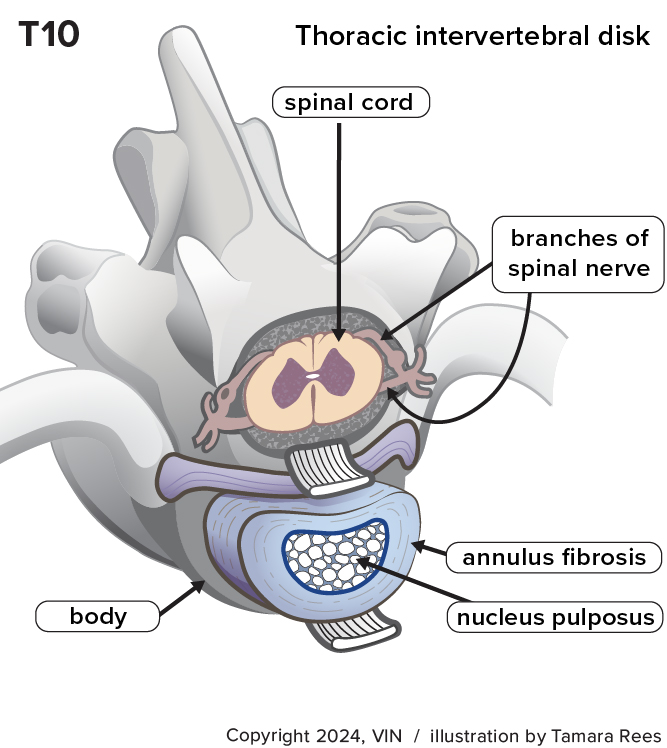
Cross-section of canine thoracic (middle back) vertebra.
Introduction
The canine spine is a complex structure composed of vertebrae, joints, ligaments, muscles, and intervertebral discs (IVDs). These discs, located between vertebrae, play a crucial role in providing flexibility and shock absorption.
Intervertebral discs have two internal parts called the annulus fibrosus, a tougher, fibrous ring, and the jelly-like inner nucleus pulposus, which act as cushions and shock absorbers of the spine.
Intervertebral disc disease (IVDD) is a disease affecting the intervertebral discs. Discs are prone to degeneration. Most degenerative disc disease is related to aging.
Degenerative Disorders
Degenerative disc diseases can lead to various conditions.
Fibroid metaplasia: This involves the nucleus pulposus losing water content, causing discs to stiffen. These degenerations may appear on imaging but usually don't cause issues. The degenerative process is usually asymptomatic (without signs) because there are no sensory nerves in the discs. As the IVDs lose their ability to act as shock absorbers, there is increased movement between the adjacent vertebral bones. This can result in the production of bony spurs (spondyle) that might join and partially fuse the vertebrae (spondylosis). This is usually asymptomatic but will be visible on X-rays.
Chondroid degeneration is genetic and starts early in life. Studies have shown that about 90% of breeds like Dachshunds and Shih Tzus will have evidence of this form of IVDD by the time they reach one to two years of age. This condition may involve the build-up of mineral deposits in discs, visible on X-rays. In most cases, it doesn’t cause pain or other issues.
02365_p_12aj5cn2gx4091.jpg
Herniation
IVDD signs surface when discs herniate (bulge) or move from their normal positions and press against or impact the spinal cord or the nerve roots.
A herniation is more common with discs that have already undergone some degeneration. Disc herniations are more likely to cause neurological deficits rather than just pain. Herniations in the thoracic spine are uncommon due to limited motion and strong ligaments. Humans with such symptoms refer to this as a slipped disc. The technical term for a slipped disc is intervertebral disc herniation (IVDH).
Because the nucleus pulposus is closer to the top of each disc and the annulus fibrosus is thinner there, disc herniations usually occur toward the top. This is where the spinal cord and nerve roots are inside the bony tube or vertebral canal.
Three major forms of such herniation are recognized.
- Disc protrusion: A lump of disc presses into the nerve roots or spinal cord.
- Disc extrusion: A portion of the nucleus pulposus ruptures through the annulus, creating a more substantial mass that affects the nerve roots or spinal cord.
- Acute Non-compressive Nucleus Pulposus Extrusion (ANNPE): A less degenerative, still gelatinous nucleus explosively ruptures through the annulus, affecting or even penetrating the spinal cord with high force. The gelatinous disc itself then mostly dissipates.
Protrusions, extrusions, and ANNPE usually occur spontaneously, with no specific event or cause preceding the onset of signs. It is common for dog owners to report that signs began after their dog fell or jumped from furniture or downstairs. In fact, disc herniation likely occurred first, resulting in the fall. The rarest form of IVDH is traumatic disc extrusion, caused by severe external trauma, such as a car accident or falling from a great height.
Symptoms of IVDH may come on gradually or very suddenly, depending on the amount of herniated disc material and the rate at which it protrudes or extrudes. Most severe signs, especially if they come on quickly, are associated with disc extrusions rather than protrusions and usually require surgery for treatment. Dogs with less severe signs need to be watched very carefully for evidence of progressive worsening since that will affect the type of treatment used.
Root Signatures
Discs do not have pain sensors, so there are usually no signs of disc degeneration. Signs of IVDD usually only occur when a protrusion or extrusion occurs. The most common sign is pain, usually due to pressure from the herniated disc.
If disc herniation occurs in the mid-lumbar and low lumbar area (or lumbosacral area, the disc between the 7th lumbar vertebra and the sacrum), signs can be related to the limb served by the affected nerve root(s). In humans, this is reported as numbness, tingling, shooting pains, and weakness in the leg. Dogs may limp, hold the leg up, or lick at the limb. This is termed a “root signature”.
Signs of Herniation
Pain is a prominent sign as well as a reluctance to walk, altered play behavior, or the inability to control the bladder or bowels.
Dogs may show behaviors like tensing stomach muscles and root signatures. These signs may include limping, holding a leg up, or licking at the limb when a nerve is compressed, as mentioned above.
Signs of mid-to-lower back herniation can include a reluctance to walk, altered play, restlessness, hunched back, vocalization, and potential tail issues.
Severity of Signs
The severity of signs depends on factors like spinal cord compression and the speed of disc herniation. Signs can range from mild pain and coordination problems to severe difficulties in standing and walking, weakness, and even paralysis.
Complications
Complications include ascending or descending myelomalacia, a rare condition where damage from a herniated disc spreads along the spinal cord. Unfortunately, this can lead to severe symptoms, and euthanasia may be recommended.
Conclusion
Understanding IVDD in dogs involves recognizing degenerative and herniation-related issues. While degenerative changes may not be painful, herniations can cause pain and loss of nerve function. Diagnostic imaging is crucial for correct diagnosis and appropriate treatment.
For more information, please see: Thoracolumbar to Tail IVDD in Dogs: Diagnosis and Treatment