Canine ear anatomy close up
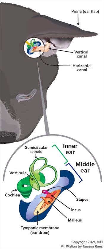
Graphic by Tamara Rees/VIN
The word otitis refers to inflammation and/or infection of the ear. Otitis media involves the middle ear (the tympanum/tympanic membrane, tympanic osseous bulla, and auditory (eustachian) tube), whereas otitis externa begins in the external ear canal. Some nerves are closely associated with the middle ear, so infection there can damage them, causing the neurologic signs often seen with middle ear infection: a head tilt, lack of balance, nystagmus (back-and-forth eye movements). These symptoms are called vestibular signs. Middle ear infections can also cause paralysis of the facial nerve, leading to a slack-jawed appearance on that side of the face.
Otitis media most often develops after an existing otitis externa travels from the external ear canal through the tympanic membrane and into the middle ear. The infection in the external ear canal leads to inflammation and damage to the ear canal and tympanic membrane, allowing the infection to enter the middle ear.
Otitis media has been found in 16% of dogs that have acute (rapid onset) otitis externa and in 52% of dogs that have chronic (long-term) otitis externa. In contrast, 63% of cats that had otitis media/interna did not have a previous history of ear infection. This is why keeping your pet's ears clean and watching for infection are important to your pet's health.
Other causes of otitis media can include infections in the nose and throat, trauma, foreign bodies, fungal infections, inflammatory polyps, cancer, etc. In addition, developmental abnormalities of the external ear canal and pharynx can lead to otitis media.
Dogs that have long, pendulous ears, such as beagles and basset hounds, are more likely to develop otitis externa, which can lead to otitis media.
The Cavalier King Charles Spaniel breed is known to have a condition called primary secretory otitis media (PSOM). Dogs with PSOM typically have mild to moderate pain in the head/neck, neurologic signs (e.g., ataxia, facial paralysis, head tilt), and itchy ears, but without otitis externa.
Diagnosis
Diagnosis includes physical examination, ear examination with an otoscope, bacterial/fungal culture, radiography, and possibly advanced imaging.
Physical Examination
During the physical examination, your veterinarian will be looking for some of these signs: head shaking, pawing at the ear, pain when opening the mouth, signs of otitis externa (ear odor, redness, etc.), head tilt, facial nerve signs, Horner syndrome, ataxia, etc. Not every affected pet will exhibit all of these signs. In addition, cats are more likely to have neurologic signs than dogs are.
Ear Examination (Otoscopy)
To do a complete examination of the ear, a deep ear cleaning may be necessary. To keep your pet comfortable, your veterinarian may decide to sedate or anesthetize your pet for the procedure. An otoscope is used to see the inside of the ear and the eardrum.
Culture and Sensitivity
Your veterinarian may take samples of the material in the ear and culture those samples to determine what medications will work best.
Radiographs
Your veterinarian may take radiographs (X-rays). Radiographs can help show changes (thickening, destruction) to the bony structures of the ear. However, even if the radiographs appear normal, that does not mean that your pet does not have otitis media.
Advanced Imaging
In some cases, your veterinarian may suggest using computed tomography (CT) and/or magnetic resonance imaging (MRI) to further evaluate the tympanic bulla. CT is usually better than MRI at detecting bony changes, while MRI is better at detecting soft tissue abnormalities.
Treatment
Cleaning both the external and middle ear canals is an important step in treating otitis media. Debris can inactivate some topical medications and prevent medications from contacting inflamed or infected tissues. Anesthesia will most likely be necessary to do the deep ear cleaning and ear flushing to remove the debris.
Your veterinarian may need to infuse (fill) the ear with medication to deliver a high concentration of antimicrobial products or corticosteroids directly to the affected area.
Topical medications may need to be used to control an otitis externa.
Systemic therapy with antibiotics, antifungals, and corticosteroids may also be used, depending on the infection.
If severe, irreversible changes to the external ear canal are found, if there are masses in the inner ear, or if medical therapy has failed to resolve the otitis media, your veterinarian may advise surgery, such as an ear canal ablation. This surgery is not undertaken lightly, but it may be necessary to control the infection. An ear canal ablation involves removing the vertical and horizontal portions of the ear canal, along with the bones of the middle ear and eardrum.
Monitoring (Rechecks)
During treatment, your veterinarian will usually want to recheck your pet's external ear canal and tympanic membrane every 10-14 days. (A ruptured tympanic membrane will generally begin to heal within 21-35 days.) Topical medications may be given until the external ear canal and the tympanic membrane are normal, and until cytology of the ear canal reveals no infection. Systemic therapy is typically given for at least 4-6 weeks.
Prognosis
In general, the prognosis for dogs is good if appropriate therapy is started in time. The prognosis is not as good if the infection is resistant to the medications; if the concurrent otitis externa is not managed adequately; if there is significant bone infection; or if there is no response to surgery. In addition, some neurologic signs (e.g., facial nerve problems, Horner syndrome) may be permanent.
Reminder
Keeping your pet’s ears clean is critical to preventing ear inflammation or infections. However, it is not always possible to prevent in some pets.