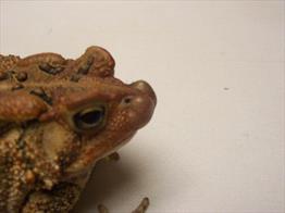
Toad with Mycobateriosis on Nose

Nodules are characteristic of bumps caused by mycobacteria in amphibians. Photo by Dr. Rick Krason.
Mycobacteriosis, often called myco or tuberculosis, refers to an infection with a species of bacteria of the genus Mycobacterium. A large number of mycobacterial species have been identified as causing disease in amphibians. Those species are termed, as a group, atypical Mycobacteria, and include Mycobacterium avium, M. fortuitum, M. marinum, and M. xenopi. These species are zoonotic, and humans may become infected from their amphibians.
Determining the type of mycobacteria is important because the type of antibiotic needed to treat different amphibian species (note: this is referring to the different species of mycobacteria that often need to be treated with different antibiotics depending on the mycobacterial species causing the disease) varies and the risk of a human getting the disease differs by species (differs by species of mycobacteria).
How do you know your amphibian may have mycobacteriosis? This can be difficult to answer as the signs of disease are most often not very specific or clear, commonly showing up as nonspecific illness, respiratory disease, anorexia or weight loss despite a good appetite.
Mycobacteria commonly cause nodules (lumps, bumps) and with some species, non-healing open wounds or ulcers. These nodules are called granulomas. They can be found in any tissue or organ in an amphibian with mycobacterial disease, but commonly are on the skin (especially the feet and toes) and can be gray, white or tan. Some of the more common tissues affected are lung, skin, alimentary tract, heart, and spleen, and so the signs of illness in your amphibian can vary depending on which tissues are diseased. Mycobacteriosis is typically acquired through an open wound or by ingesting Mycobacterium organisms. The lumps and bumps don't seem to be painful.
Mycobacteria are found everywhere in the world and they pose a significant zoonotic risk to humans, especially immune-compromised humans. Infected people commonly have open wounds on the skin of their hands. As mentioned above, the zoonotic risk to humans varies with the species of Mycobacteria. For instance, of the species reported in people from infections of amphibians, M. marinum seem to have the most significant zoonotic risk to humans, while other species have not been found to cause disease in immune-competent humans. However, all Mycobacterium spp. should be considered as a risk for immune-compromised humans.
Affected Reptiles
Mycobacteriosis can cause disease in all amphibians but, a high incidence has been reported in clawed frogs, Xenopus spp. Because most species of mycobacteria are slow growing, adult amphibians are more commonly seen with the disease. Just like for humans, amphibians that are immune suppressed are at greater risk for getting the disease and the most common cause of immune suppression in captive amphibians is incorrect or poor husbandry conditions.
Diagnosis
To diagnose illness caused by mycobacteriosis, your veterinarian will begin with a thorough husbandry history and physical examination. Commonly, your veterinarian will use a small light source shined through the amphibian, called transillumination, to detect nodules on the organs inside the body. In some cases, your veterinarian may want to collect blood and take a set of x-rays. As mentioned above, the physical exam findings are often non-specific but if your amphibian has lumps, bumps or non-healing ulcers, those signs will help to make a mycobacterial disease a possibility. Amphibians with mycobacterial disease will often show a high white blood cell count. However, because there are other causes for granulomas, lumps and bumps such as chromomycosis, nodules caused by worms under the skin, and some cancers, your veterinarian will likely suggest some additional testing to confirm the cause.
Additional tests might include a biopsy of one of the lumps, which will be sent to a lab to look for mycobacteria in the tissue. This is done with a stain; you might hear your veterinarian call it an acid-fast stain. This stain coats the bacteria and makes it easier to see.
A polymerase chain reaction (PCR) test on the tissue may also be suggested; this test is more sensitive than biopsy for detection of mycobacterial organisms. Because of the differences in how mycobacteria cause disease, react to antibiotics, and the different zoonotic potential of the various mycobacteria, your veterinarian will talk about the importance of identifying the species of mycobacteria. This can be done in an outside lab using PCR, culture and sensitivity. The culture and sensitivity are important to guide treatment by indicating which antibiotic to use.
Treatment
If, and I do mean IF, you decide to treat (most get euthanized, but the real reason for the caution in treating is the zoonotic potential and it depends on if the owners want that to take that risk) the goal of therapy is to eradicate the organism. However, before treatment is initiated, the species of mycobacteria should be identified to determine zoonotic potential to the owners and to be able to choose the correct antibiotic. the goal of therapy is to eradicate the organism. However, before treatment is initiated, the species of Mycobacteria should be identified to determine zoonotic potential to the owners and to be able to choose the correct antibiotic.
If the species identified has a high potential for zoonotic disease (such as Mycobacterium avium), your veterinarian will discuss the option of euthanasia. If treatment is decided upon, your veterinarian will discuss and make sure that you understand the zoonotic risks, expense, and are committed to the long-term therapy necessary. If you are not committed after this discussion, then euthanasia should be considered.
Antibiotic medications for treatment of most mycobacterial diseases involve the use of more than one antibiotic at the same time for at least 6 months to a year. This is because studies have shown that using only one antibiotic or treating for fewer than 6 months is likely to result in treatment failure and antibiotic resistance.
Because immune suppressed amphibians are more susceptible to mycobacterial infections and also will not be able to fight off the disease during treatment, any husbandry deficiencies discussed with your veterinarian should be corrected.
Before treatment is stopped, your veterinarian will want to repeat a biopsy of affected tissues to determine whether therapy needs to be continued. In addition, your veterinarian may want to perform physical examinations of the amphibian to look for signs that the disease has come back.
Prognosis and Prevention
The prognosis for animals with mycobacteriosis varies depending on affected tissues and mycobacterial species. Depending on the species, diagnosed treatment can range from ineffective to very effective, so identification is critical. Unfortunately, most amphibians die within 90 days of a diagnosis of mycobacteriosis.
Maintenance of a closed group with limited movement of animals into and out of the collection and having a good quarantine program are the most effective means of prevention. Quarantine of 180 days or longer may be needed to successfully detect amphibians infected with mycobacteriosis before they are introduced to the established collection.
Good husbandry practices are also important, especially hygiene practices. Atypical mycobacteria are part of the normal environment of an amphibian tank, and infection is typically the result of poor husbandry practices in the home or prior to acquiring the amphibian. Examine cage surfaces closely to ensure that they are not causing abrasions and cuts in the skin, which would promote infection. Disinfect tools appropriately between uses in amphibian cages (ask your veterinarian about the best methods). Wear disposable gloves and exchanging used gloves for new ones before touching the next cage.
Many Mycobacterium spp. are stable in the environment, especially in aquatic or moist environments. Disinfectants to use should be labeled as having effective mycobactericidal activity; the types that are available can be discussed with your veterinarian.