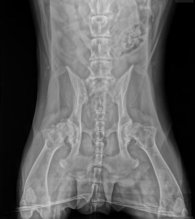
Hip dysplasia is a common condition of large breed dogs. Many dog owners have heard of it, but anyone owning a large breed dog or considering a large breed dog should become familiar with this condition. The larger the dog, the more likely the development of this problem becomes, particularly as the dog ages and loss of mobility/arthritis pain become important life quality issues.
What is Hip Dysplasia?

Radiograph of normal hips; the femoral head fits snugly inside the acetabulum, Photo by Dr. Greg Harasen
The term dysplasia means abnormal growth, thus hip dysplasia means abnormal growth or development of the hips. Hip dysplasia occurs during a puppy's growing phase, usually a large breed puppy, and essentially refers to a poor fit of the ball and socket nature of the hip. The normal hip consists of the femoral head, which is round like a ball and connects the femur to the pelvis; the acetabulum, which is the socket of the pelvis; and the fibrous joint capsule and lubricating fluid that make up the joint. The bones (femoral head and acetabulum) are coated with smooth cartilage so that motion is nearly frictionless and the bones glide smoothly across each other's surface.
See more detail on the structures of the normal joint.
When a dog has hip dysplasia, the ball and socket do not fit smoothly. The socket is flattened and the ball is not held tightly in place, thus allowing for some slipping. This makes for an unstable joint and the body’s attempts to stabilize the joint only end up yielding arthritis.

Radiograph of dog with hip dysplasia. Photo by Dr. Martha Broda
If this Disease Starts in Puppyhood why are most Affected Dogs Elderly?
Actually, there are two sets of patients typically affected by hip dysplasia. The first affected group is adolescent dogs, typically six to 18 months of age. Image 2 in this article shows the hips of such a patient. This dog has hip dysplasia but has not yet developed arthritis. Note the shallow hip sockets. Dogs in this group are commonly brought in for signs of discomfort. Radiographs are then taken and hip dysplasia is discovered.
Many young dogs with similar radiographs will not be in pain and thus will not end up coming in for an evaluation. As they age, they develop bony spurs along the margin of the socket, mineralization of the joint capsule, cartilage wear, and inflammatory change in the joint (i.e. degenerative arthritis) and become painful and now these dogs come to the veterinarian for an evaluation.
Why Are There Differences in Age and Size for Patients with Hip Dysplasia?
Obviously, different individuals may have different degrees of dysplasia. A dog's weight makes a difference (a lighter dog can more easily tolerate a more abnormal hip joint). The muscle mass supporting the joint is greater in a younger dog and helps reduce the stress directly on the bones. Still, some dogs have truly shocking radiographs and virtually no symptoms while others show relatively subtle changes and are very uncomfortable. It is not known why there is not a better correlation between radiographs and actual pain.
How Can Owners Tell if Their Dog is Having Discomfort?
Do not expect a dog with dysplasia (or any other chronically painful condition for that matter) to cry or whine in pain. Instead, discomfort is shown with reduced activity, and difficulty rising or lying down, or going upstairs. A characteristic swivel of the hips is seen from behind and classically stairs are taken in a bunny hop fashion.
What Causes Hip Dysplasia?
The primary cause of hip dysplasia is genetic but inheritance of this trait is not as simple as a dominance/recessive relationship like we study in high school biology. Normal dogs can breed and yield dysplastic offspring as the condition may skip generations. Furthermore, dogs with a genetic picture conducive to hip dysplasia still must contend with other factors such as level of exercise at an early age, nutritional factors, hormonal/neutering factors, and other environmental situations.
Preventing hip dysplasia primarily focuses on breeding dogs with normal hips. The problem with this approach is that dogs often do not develop signs of hip dysplasia until well after they have been bred. A genetic test would be of great value in dog breeding, but currently, there is only such a test available for Labrador retrievers; identifying dogs with less than stellar hip quality so as to exclude them from breeding is done via OFA and PennHip certification (see sections on registration below).
It is important to consider the breed of dog when choosing a puppy for many reasons, including potential genetic orthopedic problems. Hip dysplasia is typically a problem for large, stocky dog breeds. Small dogs and lean, slender breeds such as sighthounds rarely develop hip dysplasia. If you have settled on a breed that has an issue with hip dysplasia, be aware of the certification process of the parents. The Orthopedic Foundation for Animals publishes statistics on affected breeds.
Other than selective breeding, it is possible to manage other factors in hip dysplasia development when raising a predisposed puppy that may help make the genetic issues less severe.
Nutrition
Nutritional factors are important in developing hip dysplasia. For example, it has been popular to try to nutritionally push a large breed puppy to grow faster or larger by providing extra protein, more calcium, or even just extra food. Practices such as these have been disastrous, leading to bones and muscles growing at different rates and creating assorted joint diseases of which hip dysplasia is one. One study showed that when puppies of hip dysplasia-prone breeds were allowed to free feed, two-thirds went on to develop hip dysplasia while only one-third developed it when the same diet was fed in meals. Another study showed German Shepherds were nearly twice as likely to develop hip dysplasia if their adult weights were above average. Studies such as these have led to puppy foods designed for large-breed puppies, where the optimal nutritional plane is lower than for small-breed puppies. After puppyhood, maintaining a lean body condition seems to be helpful in lessening arthritis signs.
Exercise
Exactly what the proper preventive exercise regimen might be is yet undefined. One study showed that puppies had an increased risk for hip dysplasia if they were allowed to freely run up and down stairs before age three months. The decreased risk was found in puppies allowed off-leash exercise, such as in the backyard, before age three months.
Neutering Age
There is some controversy about the effect of spaying/neutering before puberty. Male dogs rely on testosterone to stop bone growth and early neutered males will grow taller as their bones grow for a longer period if testosterone is removed early. This may lead to a predisposing disparity in the growth of bone and muscle. There appear to be predisposing factors for early spay as well, assuming the dog has the genetic predisposition. How much added predisposition comes with early spay/neuter and for which breeds remains controversial. In one study, it was found that neutering before age 5.5 months was associated with a 6.7% incidence of hip dysplasia while neutering after age 5.5 months was associated with a 4.7% incidence of hip dysplasia.
Severe hip dysplasia

Radiograph showing severe hip dysplasia. Photo by MarVistaVet
How Can I Find Out if My Dog Has Hip Dysplasia?
There are two reasons to pursue testing: to explain a dog's discomfort/rear weakness or to screen a dog for breeding purposes. If a dog is not going to be bred and is not in any apparent discomfort, there may be no benefit to looking at the conformation of the bones in a radiograph except possibly to look back at a future time to get a sense for the progression of bony changes.

By MarVistaVet
The first step in diagnosis is an examination. Your veterinarian will likely extend the dog's hind leg backward to check for pain as hip dysplasia causes pain on hip extension. The dog may be asked to walk around to demonstrate the possible hip swivel. Another test involves having the dog lie on its back with a hind leg perpendicular to the body. As the leg is moved away from perpendicular to the body, a dysplastic hip will generate a pop as the femoral head slips to the center of the acetabulum. This pop, which can be felt if your hand is resting on the hip during the exercise, is called an Ortolani sign. You may hear this term used as hip dysplasia is discussed.
The true confirmation of hip dysplasia comes with radiography. The dog must be radiographed on their back with both legs positioned straight down. This posture is painful to a dog with dysplasia so to get maximum cooperation from and comfort for the patient, sedation is needed. The seating of the femoral heads in the acetabular sockets is examined and assessed for arthritis.
Above are two radiographs: normal hips in Image 1 and dysplastic hips in Image 2 (and severely dysplastic hips in Image 3). It is easy to see how the femoral heads are not well seated in their sockets in the dysplastic hips. This puppy in the above left has a subluxation, which means the hips are nearly out of the socket completely. Over time, the femoral heads will flatten and the sockets will become even more shallow. Bone spurs will develop around the joint capsule as the body attempts to stabilize the abnormal joint.
What is OFA Registration?
When purchasing a puppy, particularly one of a larger breed, often the parents will be listed as “OFA Good” or “OFA Excellent.” What this means is that the breeder has had the hips of the parent dogs certified by the Orthopedic Foundation for Animals. The OFA is an organization with the goal of reducing the incidence of hip dysplasia (though now it is also possible to obtain certification for elbows, thyroid function, and other issues). The idea here is that a dog for breeding can have radiographs taken at the age of 24 months. The radiographs are sent to the OFA for review by several independent radiologists where they are graded. Hips that are rated as “good” or “excellent” receive a registration number. Offspring of OFA-certified parents would be less likely to develop dysplasia themselves, however, it is important to realize that a dog with excellent hips at age 2 may not have such excellent hips at age 5, 7, or 10. OFA certification is no guarantee that a dog will not develop hip dysplasia symptoms in the future and does not guarantee that the offspring will not develop hip dysplasia but, as mentioned, until a DNA test for hip dysplasia is developed parental certification is the best we can do.
What is PennHip Registration?
Many people with potential breeding dogs do not want to have to wait two years for OFA registration. The University of Pennsylvania Hip Improvement Plan, developed by Dr. Gail Smith, allows for another way to predict if a dog will develop hip dysplasia. For PennHip certification, the veterinarian taking the radiographs must receive specific training and special equipment is necessary. The pet is anesthetized and two radiographs are taken: one with the femoral heads compressed (pushed into the acetabula as far as they will go) and one with the femoral heads distracted (pulled out of the acetabula as far as they will go). A measurement called a distraction index is calculated from these radiographs, the idea being that a tighter-fitting hip - one allowing less distraction - is less likely to develop dysplasia. Each dog breed has a different range of distraction that is considered acceptable. Puppies can be certified as young as 16 weeks of age with this system.
Is Surgery the Best Treatment for Hip Dysplasia?
There are many surgical options for hip dysplasia and it is important to understand which patients benefit from which surgery. Some surgical procedures are controversial and some are not. All will entail a recovery period as well as expenses. Both hips need not necessarily be treated surgically; treating one hip is often enough to yield good results. Hip surgery is relatively expensive and a surgery specialist may be required, depending on the procedure. If you are considering surgery for your dog, these are the procedures to know about.
Femoral Head/Neck Ostectomy
This surgery is commonly referred to as the Femoral Head/Neck Ostectomy(FHO) and is best used for smaller dogs (50 lbs/22.7kg or less) or very active dogs. Here, the femoral head is cut off and removed, allowing the joint to heal as a false joint (just a capsule connecting the two bones but no actual bone-to-bone contact). If the dog is not carrying too much weight, a false joint is strong enough. If the dog is very active, a false joint will form quickly. The pet typically does not want to use the leg for the first two weeks but should at least be partially using the leg after four to six weeks. The leg should be used nearly normally after a couple of months. Many veterinarians are well experienced with this surgery and often a specialist is not needed. This surgery is typically substantially less expensive than the other procedures.
Triple Pelvic Osteotomy
This surgery is appropriate for young (8-18 months) dogs with dysplasia but without degenerative arthritis changes. This means that there is a window of opportunity for this surgery and if the dog develops arthritis or becomes too old, it will be too late to do this surgery. Here, the ill-fitting acetabulum is essentially sawed free of the rest of the pelvis, re-positioned for a tighter fit on the femoral head, and then plated back into place.
Many times surgery on one hip leads to positive changes in the other hip so surgery on the second hip is not necessary. Alternatively, it is possible to do the TPO on both hips if it seems clear that ultimately both will need surgical correction. Many general practitioners will not feel comfortable doing this procedure, so discuss with your veterinarian whether a referral to a board-certified surgeon or a surgeon with extensive orthopedic experience is in your pet's best interest. Aftercare involves a good three to four months of exercise restriction. No leashed walks, except to go outside for elimination, for two months.
The biggest problem with the TPO is determining whether the patient really needs it. If the dog is not experiencing hip discomfort at the time of diagnosis, he may not experience hip discomfort until he is elderly, and should he really have an invasive procedure early as a preventive? On the other hand, if he is having discomfort at a young age, the arthritis is likely to only get worse and early surgery could help this tremendously. These are matters to discuss with the surgeon.
Total Hip Replacement
This procedure is for dogs with established degenerative hip changes. For these dogs, the best choice may be to simply replace the hip or hips with a prosthetic hip. This procedure may sound radical but it has been commonly performed for over 30 years in dogs with great success. It is a highly invasive procedure, obviously, and infection must be avoided at all costs (no skin disease can be in the skin over the hips, extra precautions for sterility are used). In other words, when complications occur they have the potential to be severe. Complications have about a 10 percent incidence. Expect about three months of exercise restriction after this procedure. Usually, only one hip receives surgery at a time. Often only one replacement is needed and the pet does well enough not to need surgery on the other side.
Juvenile Pubic Symphysiodesis
This surgery is performed on young puppies before age five months, so it is generally done as a preventive procedure before it is known if the puppy will indeed have dysplastic hips but after hip laxity has been detected. The pubic symphysis is the cartilage seam connecting the right side of the pelvis to the left side. As an individual matures, this cartilage converts to bone and the two halves of the pelvis fuse permanently. This surgery prematurely seals the symphysis, which in turn results in rotating the developing hip sockets into a more normal alignment. This is the same re-configuring as the TPO above but instead of doing the reconfiguring with surgery, it is done by directing the puppy’s own bone growth. The window is very narrow for this procedure so if a puppy is a predisposed breed it may be worth getting screening radiographs around age 4 months even though no physical discomfort has developed. It may be worth having this preventive procedure while the puppy is still at an age to benefit from it.
No matter which, if any, procedure is selected, it is important to get an idea of expenses, recovery procedure, and what is entailed from your veterinarian and the surgeon before making a decision.
What Non-Surgical Treatment is Available?
Non-surgical treatment of hip dysplasia is essentially the same as a non-surgical treatment for any other type of arthritis. There are nutritional supplements to help repair cartilage, pain medications, and anti-inflammatory medications. Physical therapy and massage are also important and helpful in non-surgical joint therapy. There is no medical way to reverse or prevent hip dysplasia; the medications described are for pain management. For details, see medications for degenerative arthritis.