Leptospira spirochetes
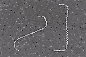
Photo by Janice Haney Carr, courtesy of CDC
About the Organism
Leptospira organisms are spiral-shaped bacteria called spirochetes. There are several species of leptospires, but the ones that cause disease have been grouped into one particular species called Leptospira interrogans sensu lato. From here, Leptospira interrogans sensu lato has been sub-classified into smaller related groups called serovars. Over 250 serovars have been named and at least 10 are important for pets. A vaccine for dogs, however, exists against only four serovars. Different serovars produce different types of disease and are in different geographical areas.
Canicola: This serovar is the most common in Mexico. It primarily produces kidney disease. Watch out for this serovar in states near the Mexican border.
Pomona: This serovar is associated with livestock and tends to produce severe kidney and liver disease.
Grippotyphosa: In a survey of over 1,200 healthy dogs in Michigan, 24% tested as exposed to Leptospira, and the Grippotyphosa serovar was the most common.
Icterohaemorrhagiae: This serovar is mostly associated with exposure to rats and rat urine in standing water. It tends to attack the liver.
Leptospires live best in warm, slow-moving water, such as after heavy rains or flooding. After the water clears, they contaminate the soil for many months. Wildlife are common carriers of infection. A survey in Connecticut found 36% of raccoons had been exposed, while a survey in Illinois found 48% of raccoons had been. Another survey found that 50% of rats had been exposed. Classically, infection of dogs and humans stems from the urine of infected animals getting into environmental water. Leptospirosis is a common human disease in tropical areas, especially where rice is farmed, and rats infest the paddies. Health authorities believe that rat populations are involved in the rising incidence of canine Leptospirosis in urban areas, and Leptospirosis is no longer considered a rural disease. Leptospires can survive for months in contaminated soil.
This all sounds outdoorsy, but the 2021 Leptospirosis outbreak in Los Angeles started in boarding facilities where dogs are commonly exposed to the urine of other dogs in play yards and kennel drainage pathways. Anywhere with potential urine contact is a potential home for Leptospira organisms.
Canine Infection
Dogs become infected by leptospires when irritated or cut skin comes into contact with infected urine or water contaminated with infected urine. Alternatively, bite wounds, exposure to reproductive secretions, and even eating infected tissues can transmit this infection. The organisms quickly spread through the bloodstream, leading first to inflaming the blood vessels: fever, abnormal bleeding, abnormal bruising, and tissue edema appear after an approximately seven-day incubation period. By two weeks post-infection, the leptospires have set up shop in the kidneys, where they continue to generate inflammation, pain, and potentially total kidney failure and the inability to produce urine. Some serovars also go to the liver and generate inflammation there, though the liver disease is generally not as severe as that of the kidney.
A particularly devastating situation occurs if the organism gets into the lungs where the leptospire toxins produce what is termed “Leptospira Pulmonary Hemorrhage Syndrome." The lung bleeding that results is associated with a 70% mortality rate and bodes especially poorly.
If the dog is able to keep the acute illness at bay, a chronic form may emerge. There can be more chronic kidney insufficiency and/or hepatitis. Furthermore, long-term immune stimulation can lead to a deep eye inflammation called uveitis that can cause the eyes to look cloudy or even change color. If the disease is treated in this form, it may not be possible to reverse the long-term damage that has already set in.
The Different Clinical Pictures of Leptospirosis
As you might gather, Leptospirosis can look different: fever with bruising and bleeding; fever with different degrees of kidney failure; liver disease and kidney disease together; chronic hepatitis; and eye inflammation, etc. Another way to look at Leptospirosis is to look at the time frame of the illness and how quickly or slowly it came on.
Peracute Disease: Peracute disease means very sudden onset. These are usually younger dogs with an overwhelming exposure. The large amount of leptospire toxin causes rapid death before the kidney or liver disease even happens.
Acute Disease and Subacute Disease: This is more of the classic form described above—fever with bruising and bleeding, general muscle pain, and a painful belly from kidney and/or liver disease. There may be jaundice and inflammation in the eyes that makes them look cloudy.
Chronic Disease: Recurring fevers, chronic hepatitis, chronic kidney disease, uveitis, poor appetite, weight loss.
Younger dogs (less than one year of age) tend to get the most severe forms of Leptospirosis.
87-100% of infected dogs will have some degree of azotemia, which means kidney values will be elevated on routine blood testing.
Excessive water consumption is frequently seen at home when this happens.
Testing
PCR Testing
PCR testing is used to detect leptospire DNA. A blood sample is best in the first 10 days after infection, but after that, a urine sample is more likely to be positive. It may be prudent to submit both blood and urine samples. Past vaccination will not interfere with this test, although antibiotic exposure certainly will, and results can be back in a matter of days. PCR testing will not determine which serovar is present.
MAT Testing (the Traditional Test)
The Microscopic Agglutination Test, or MAT titer, is still considered the test of choice, though it has some disadvantages. It measures antibody levels against different L serovars, with the idea that the one with the highest level is most likely the serovar causing the disease.
Antibody levels are expressed as titers, which are ratios reflecting how much dilution is needed before it is too diluted to detect antibodies. For example, a titer of 1:32 means a serum diluted out 32 times still had detectable antibodies. A titer of 1:32 may sound high but it is actually low; an MAT titer must be at least 1:800 to be considered positive. If the serovar under consideration is one that might have been included in a past vaccination, the titer must be higher (1:3200) to be considered positive.
To really obtain high confidence in the diagnosis of Leptospirosis, a second titer is submitted two to four weeks later, showing at least a four-fold increase in antibody production. Treatment with antibiotics should not interfere with the validity of the second (or "convalescent") titer level.
There are two problems with this testing:
- No one wants to wait two to four weeks to confirm the diagnosis, especially with a disease contagious to humans. PCR testing gets results much faster.
- Vaccination interferes with results (remember, the entire goal of vaccination is to generate an antibody titer). Vaccination history can make interpretation difficult.
In-House Test Kits and Other General Antibody ELISA Tests
Recently in-house screening tests have become available so that a result can be obtained in 20 minutes or so. These tests screen for antibodies against Leptospira organisms. They are either positive or negative. They will not tell you which serovar is involved nor how high the titer is. They will not distinguish antibodies from vaccination versus those from true infection. Clearly, a positive test needs to be followed by another test. A negative test, however, is very helpful (see below).
Other Tests
In the past, cultures and darkfield microscopy were used to detect leptospires. This technology is now considered old-fashioned.
Which Tests to Use?
A good approach to begin with is a general antibody test combined with urine and blood PCR testing. If any PCR test detects Leptospira DNA either in blood or urine, the infection is confirmed. A MAT test will determine which serovar is afoot and the antibody level can be tracked to be sure the treatment is working.
If the PCR tests are negative (meaning Leptospira DNA was not detected) but a general antibody test is positive, the MAT test will determine if the antibodies are related to prior vaccination or active infection.
If PCR tests are negative (Leptospira DNA not found) and no antibodies are found either, then it should be safe to cross Leptospirosis off the list of possible diagnoses.
Treatment
Fortunately, Leptospira interrogans sensu lato is sensitive to doxycycline, a readily available antibiotic. Leptospires are cleared from the blood within 24 hours of starting it, but it takes about a week for them to clear from the urine, so it is important to wear gloves, goggles, etc., and be conscious of contamination during urine cleanup. Infected animals should be isolated from other animals at least until their antibiotic course is complete and probably for a couple of weeks after. Check with your veterinarian for instructions.
Intravenous fluids are crucial to support blood flow through the damaged kidneys so that recovery is possible. Any areas at home that have been contaminated with urine should be disinfected with an iodine-based product and you should wear gloves while cleaning any urine. Prognosis is guarded depending on the extent of organ damage; with appropriate treatment 80-90% survival rates are reported.
This sounds wonderful, but it is important to keep in mind factors that can interfere with this rosy outcome. While most Leptospirosis-related kidney injury responds to fluid therapy which can be given by most animal hospitals, more severe cases can require dialysis, which has limited availability. As mentioned, lung involvement has poor survivability. Infection with the Pomona serovar is associated with more severe disease.
A good two weeks of doxycycline is generally needed. If this antibiotic is not tolerated, amoxicillin may also work.
Previously infected dogs may become re-infected. Past infection does not confer future immunity.
Prevention
Vaccination against Leptospira organisms is only available for some of the serovars or varieties that we know of. Leptospira interrogans sensu lato is only available for the serovars called Canicola, Grippotyphosa, Pomona, and Icterohaemorragiae.
Some vaccines cover all four serovars, while others cover only two out of four.
Vaccination against two variants, Canicola and Icterohaemorragiae, has been traditional for dogs as it is included in the basic distemper shot (DHLPP - the “L” stands for Leptospirosis). The American Animal Hospital Association vaccine guidelines (as of 2024) recommend vaccination against Leptospirosis to be a part of the core (always recommended) vaccines for dogs and to use a vaccine covering all four serovars whenever possible. Research has shown that all dogs may be at risk of Leptospirosis regardless of age, breed, location, season, or other factors, but it is more common in warmer, humid climates with high rainfall.
As with all vaccines, there is a risk for reactions. In the past, the Leptospirosis vaccine was thought to be associated with a higher chance of immunological vaccine reactions, but vaccines made from leptospires grown in protein-free media have made vaccination reactions less likely.
Common vaccine reactions may include fever, swelling at the injection site, and joint/muscle pain. These symptoms should resolve after a day or so. Small dogs are more likely to experience reactions. Some vaccine reactions can be serious, so consider talking to your veterinarian about reactions before vaccination.
Vaccinations should reduce the severity of the disease but will not prevent infected dogs from becoming carriers.
Other important aspects of prevention include controlling rodents and other wild animals in the pet's environment and removing standing water so that your pet cannot drink it.
The Infection in Humans
The Centers for Disease Control and Prevention monitors Leptospirosis cases in people. Humans can be infected by direct contact with urine from an infected animal or food, soil, or water contaminated with urine from an infected animal. The most common species to transmit Leptospirosis to humans are dogs, rats, raccoons, skunks, opossums, and other marsupials, cows, pigs, and mice. Recreational activities involving water and exposure to flood waters are also associated with human outbreaks. Other human risk factors include farm work, animal care work, camping, and sewer work. Remember, leptospires come from contaminated urine, which, in turn, contaminates environmental water and soil.
The same symptoms occur in humans as would be seen in dogs.