
You should seek veterinary attention immediately if you see any of the following:
- Your pet squinting or protecting an eye
- Any suspected trauma to the eye
- Abnormal appearance of the eyeball
- Excessive redness to the white part of the eye (sclera)
- Any time the eyelid cannot cover the eyeball
These can indicate potentially serious eye problems that can risk your pet’s vision.
Conditions like trauma, glaucoma, perforation of the cornea (the clear membrane in the front of the eye), serious infections, foreign bodies, and autoimmune diseases can all affect your pet’s eye and may need medical care.
Take care not to get bitten when treating your pet, and use a muzzle when needed to stay safe.
ProtopsedEye
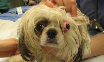
Small dog with protopsed eye (eye popped out of socket). This is an absolute emergency, and must be treated rapidly. Photo courtesy of Emergency Specialists Board.
What to Do
- If an eye has been dislocated from the socket (proptosis) or the lids cannot close over the eyeball, keep the eyeball moist with contact lens wetting solution, K-Y jelly, water, or moist compresses.
- If an irritating chemical or other product accidentally gets into the eye, flush it with running water, contact lens saline, or homemade saline solution squeezed from a compress or a sponge for a minimum of 15 minutes. (Saline: dissolve 2 teaspoons of table salt in 1 quart of water)
- Always seek veterinary attention immediately. Eyes are quite fragile and just a few minutes could mean the difference between sight and blindness. Referral to an eye specialist (ophthalmologist) may be needed for more severe cases.
What NOT to Do
- Do not attempt to treat the eyes, or remove a foreign object, yourself.
- Do not try to push a proptosed eyeball back into the socket. This must be done under anesthesia so as not to cause damage to the eyeball's interior.
Browse the complete Veterinary Partner First Aid collection.