Coccidiosis refers to an infection by one or more of the Coccidia species that infect reptiles. Coccidia are protozoa (a type of single-celled parasite) that are commonly seen in fecal samples and are often present in low numbers in healthy reptiles but can cause many different signs of illness and infection in reptiles. In one recent study that looked at 330 bearded dragon fecal samples, 49% to 65% had pinworms and 25% to 41% had coccidia. Coccidia are acquired from cage-mates or from the environment of the infected reptile; crickets may be vectors (spreaders) but are not the source of infection. Reptiles with untreated coccidia infection may stop eating and appear listless; these animals will not appear healthy or might begin to have a thin, malnourished appearance.
These parasites can infect both the intestines (intestinal) and the tissues (extra-intestinal) of reptiles. A recent study showed coccidia initially infect the upper small intestine, then over time spread down the GI tract to the colon or large intestine. Signs associated with intestinal coccidiosis may include poor growth, weight loss, bloody feces (melena), and diarrhea. Signs of extra-intestinal coccidiosis vary according to the infected tissue and may include sudden death, sore nose, lack of appetite, depression, and reluctance to move. Often, no signs of a problem are seen, and coccidia are detected during a routine fecal examination.
Coccidia are generally stable in the environment and are passed in the feces by infected reptiles. This is known as a fecal-oral mode of transmission, which is the most common route of infection for coccidia with direct life cycles (this is when the reptile can be infected by the coccidia in the cage). For coccidia with indirect life cycles (this means the reptile cannot be directly infected with that coccidia species), definitive hosts are typically infected by ingesting intermediate hosts such as insects.
Three families of coccidia are found in reptiles: Cryptosporidae (causes Cryptosporiosis), which is discussed in a separate section; Eimeriidae, of which the genera Caryospora, Eimeria, and Isospora have been described in reptiles; and Sarcocystidae, of which the genera Besnoitia and Sarcocystis have been described in reptiles. Eimeria spp. are the most commonly described coccidian parasites of reptiles. Eimeria have a direct life cycle and usually are found in the intestines of infected reptiles.
There have been no documented cases of zoonosis (infecting a human) with a reptile coccidia species.
Affected Species
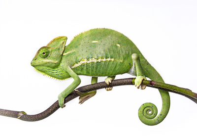
Veiled chameleon

Coccidiosis is seen in all species of reptiles that have been investigated. With intestinal Coccidia spp., young and older animals tend to be affected by the heaviest infestations and show the most significant clinical signs. No age association has been found for disease seen with extra-intestinal coccidia and no sex predilection has been noted for any reptile coccidiosis.
Young animals, animals kept in high population density, and animals kept under conditions of poor hygiene are at the greatest risk to show disease from coccidiosis. For coccidia with indirect life cycles, mixed species collections are at greater risk.
Coccidia organisms can infect many different reptile species and have been found to infect a variety of tissues; examples include:
- Tissue cysts in the kidneys of basilisks (Basilicus basilicus); intestine, liver, and spleen of ameiva (Ameiva ameiva); and heart of wall lizards (Lacerta dugesii).
- Caryospora chelonae, which has a direct life cycle, is a significant pathogen in green turtles (Chelonia mydas), causing primarily intestinal lesions, although lesions may also be in the kidney, thyroid, and brain.
- Choleoeimeria hirbayah is a significant pathogen of veiled chameleons (Chamaeleo calyptratus).
- Intranuclear coccidiosis is a significant disease of tortoises, causing high mortality:
- Organisms are found in cell nuclei of numerous organs, including GI tract, liver, kidney, and spleen.
- In Sulawesi tortoises, this organism has been associated with erosive rhinitis (nose infection). The life cycle of this organism is not known.
- Isospora jaracimrmani can be a significant pathogen in veiled chameleons (Chamaeleo calyptratus), and I. amphiboluri is the most common coccidia seen and can be a significant pathogen in bearded dragons (Pogona vitticeps).
- Klossiella boae has been reported from the kidneys of a Boa constrictor.
- Significant intestinal disease has been associated with Sarcocystis in a bull snake (Pituophis melanoleucus sayi).
Given the number of different reptile species and groups that have been found infected and diseased by coccidia, it is safe to assume that any reptile can have coccidiosis.
Diagnosis
Your veterinarian will start by taking a thorough medical history and giving a physical examination. Findings in cases of infection with coccidia that is causing disease vary according to the species of coccidia and the host; often, there are no abnormalities. Abnormalities that are commonly seen may include poor growth, weight loss, and depression. Because many diseases other than coccidiosis can cause these signs, your veterinarian may need to perform some additional tests and ask questions to get a diagnosis that may include a thorough history, taking blood for a complete blood count and plasma chemistry, X-rays, fecal examination or PCR testing. This is critical because the disease significance and approach vary greatly depending on coccidian species, so identifying the coccidia is essential.
Some species of coccidia can be seen on blood smears, so your veterinarian may want to collect blood to have a look. One of the most common ways to identify an infection with coccidia is through the feces and so a fecal floatation test may be done. Your veterinarian will look at the feces under a microscope, then determine the number of oocysts that are on the slide. Your veterinarian will decide if they are in high enough numbers to treat and what medication would work best.
A nested polymerase chain reaction (PCR) test, which screens for the genetic material of coccidia, is available for all coccidia from the University of Florida. For this test, your veterinarian may need to perform a biopsy or other procedure to collect the tissue samples needed for PCR or to have a pathologist look at them under a microscope to help them make a diagnosis.
Treatment
The goal of treatment varies according to the species of coccidia and host. For instance, for species that cause milder disease, treatment may not be indicated, especially in nonbreeding situations or where parasites have indirect life cycles. For coccidia with direct life cycles, strong hygiene practices are necessary to prevent re-infestation. This is frequently underemphasized, and without this, drug treatments will fail.
For debilitated animals, your veterinarian may want to provide supportive care such as fluid therapy, and treating a secondary infection using antibiotics may be indicated.
If a coccidia with an indirect life cycle is diagnosed, your veterinarian will discuss how to limit or remove access to intermediate hosts to prevent infection.
Your veterinarian may prescribe an anticoccidial drug for your reptile. Some of the drugs that may be considered include ponazuril (Marquis®), toltrazuril (Baycox®), nitazoxanide (Navigator®), amprolium hydrochloride, sulfadimethoxine, and trimethoprim/sulfamethoxazole.
Sulfa drugs in general seem to be poorly effective at eliminating coccidia: sulfadimethoxine administered for 21 days reduced oocyst (immature coccidia) shedding but did not eliminate coccidia in all animals. Ponazuril treatments effectively stop oocyst shedding (your veterinarian will recheck a fecal sample two weeks post-treatment to be sure). Pinworms probably are not harmful in small numbers, but in massive numbers are associated with decreased thriftiness, impaction, and cloacitis. If pinworms are also present, your veterinarian may recommend fenbendazole treatment for a number of days, then wait two–three weeks, then repeat for five days, which is often effective. (Your veterinarian will recheck a fecal sample six weeks post-treatment and again at six months.) Both coccidia and pinworms have direct fecal-oral life cycles so it is important to remove feces daily, clean the cage well, discard bedding, and scrub the ventrum (your pet’s underside, or belly) and back half of the reptile as directed by your veterinarian.
Enteric coccidia with direct life cycles should have follow-up testing every six months until they have had two years of negative fecal flotation examinations.
Prevention
Good hygiene is absolutely critical in preventing coccidia infections. Other preventive measures include maintaining a closed group, testing populations, strict quarantine, eliminating access to intermediate hosts, and the best husbandry that you can provide.
Coccidia with a direct life cycle, like those found in bearded dragons, can increase to tremendous numbers in an enclosure. The oocysts, or eggs, can survive for weeks in the environment. Fastidious, daily cleanings are necessary to remove all feces and feces-contaminated food and water. Insects and other food items must be removed on a daily basis because they are another source of contamination and reinfection.